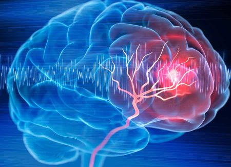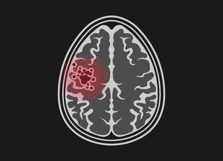
Arteriovenous malformation (AVM) refers to abnormal connections between arteries and veins in the body. This condition can replace normal tissues and affect blood flow. It is usually congenital and can cause symptoms such as headaches, neurological issues, or bleeding. Treatment, depending on the symptoms and characteristics of the AVM, may involve surgery, embolization, or radiation.
What is Arteriovenous Malformation (AVM)?
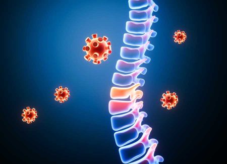
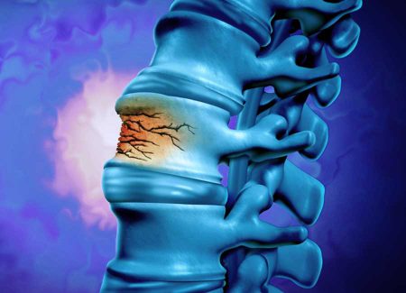
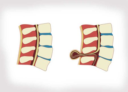
Arteriovenous malformation (AVM) is a type of vascular anomaly characterized by direct and abnormal connections between arteries and veins. This condition causes blood to bypass the capillary network and flow directly into the veins. The tangle of vessels can damage the surrounding tissues and sometimes cause harm by bleeding. Although AVMs are most commonly found in the brain and spinal cord, they can also occur in other parts of the body.
What are the Symptoms?
AVMs sometimes show no symptoms and are discovered incidentally. However, the most common symptoms of Arteriovenous Malformation, depending on their location and size, can include:
- Headaches: Severe and recurring headaches may occur.
- Seizures: Seizures may happen due to disrupted electrical activity in the brain.
- Visual Disturbances: Problems such as loss of vision may arise.
- Speech Difficulties: Issues with language and speech may occur.
- Motor and Coordination Loss: Weakness or coordination problems in the limbs may develop.
- Sensory Loss: There may be a loss of sensations such as pain or temperature.
- Bleeding: AVMs in the brain can lead to serious and potentially life-threatening hemorrhages.
What are the Causes?
The exact cause of arteriovenous malformation (AVM) is not well understood, but it is generally congenital, resulting from an abnormal process in vascular development during the embryonic period. In some cases, genetic factors may be influential, but it is still unclear what precisely causes AVM formation.
AVMs result from abnormal connections between arteries and veins, replacing the normal capillary beds and causing direct arterial to venous flow. This abnormal structure can lead to vessel dilation, thinning, and weakening.
Understanding the precise factors in AVM development is the focus of ongoing research to develop more effective treatment and prevention strategies.
Diagnosis and Tests
The main methods used for diagnosing AVM include:
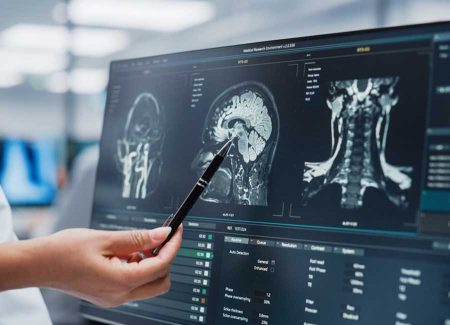
- Magnetic Resonance Imaging (MRI): Provides detailed images of AVMs in the brain and spinal cord.
- Computed Tomography (CT): Quickly detects bleeding if present.
- Angiography: Involves injecting contrast material into the blood vessels to examine the vessel structure and abnormal connections in detail. It is the gold standard for diagnosis.
- Ultrasound: Used to evaluate AVMs in other parts of the body.
Arteriovenous Malformation Treatment Methods
Observation:
For asymptomatic and low-risk small AVMs, active monitoring and regular check-ups are recommended.
Surgical Treatment:
- Resection: Complete surgical removal of the AVM is ideal for accessible AVMs causing significant symptoms.
- Endovascular Embolization: A minimally invasive procedure where embolic material is injected into the vessels feeding the AVM to reduce its size or lower bleeding risk.
Radiotherapy:
- Stereotactic Radiosurgery: Procedures like Gamma Knife use high-dose focused radiation to shrink the AVM. It is preferred for AVMs that are not suitable for surgery or are hard to reach.
Frequently Asked Questions
The recovery process varies from person to person and depends on the type of treatment, the size, and the location of the AVM. Full recovery can be achieved in some cases, while others may experience permanent neurological damage.
Treatment depends on the symptoms, characteristics, and location of the AVM. In some cases, if symptoms are minimal or absent and the AVM is small, treatment may not be necessary, and regular monitoring is advised. However, if symptoms are present or the AVM is large, treatment should be considered.
The duration of AVM treatment varies based on the treatment method, the size, and the location of the AVM. Some surgical procedures may take a few hours, while other treatment methods can take several days or weeks.
Although AVM treatment is generally successful, recurrence can occur in some cases. Therefore, regular follow-up with a doctor is important after treatment.
Yes, arteriovenous malformation (AVM) can be life-threatening. The risk depends on the size, location, and symptoms of the AVM. In some cases, the presence of an AVM can cause serious complications and pose a threat to life. Brain AVMs, in particular, carry the risk of sudden bleeding, which can lead to severe neurological damage or death. However, early diagnosis and appropriate treatment can help reduce or prevent these risks. Therefore, individuals diagnosed with an AVM should consult a specialist to evaluate the best treatment options.
Surgical methods for AVM treatment carry some risks, including bleeding, stroke, infection, neurological damage, anesthesia complications, a long recovery period, and the risk of recurrence. However, these risks are considered against the potential benefits and suitability for the patient by the doctor.
AVMs, particularly when located in critical organs like the brain, are serious conditions that require careful management. With a good treatment plan, most patients can control symptoms and prevent serious complications like bleeding, thereby improving their quality of life. Treatment should be personalized to the AVM’s characteristics and the patient’s overall health. Early diagnosis and effective treatment are crucial since each bleeding event can increase brain damage.

