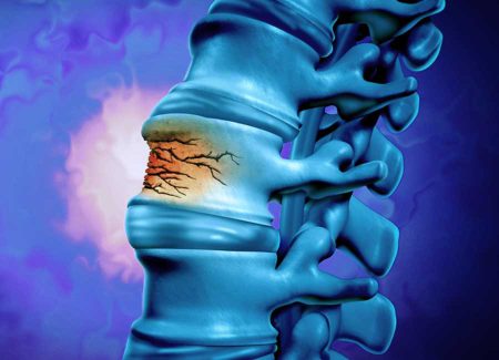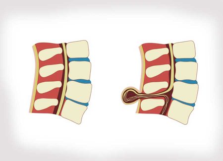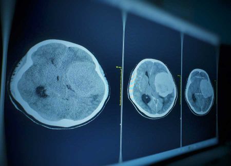
Cavernomas are vascular lesions typically found in the brain or spinal cord, usually benign in nature. These lesions, also known as cavernous angiomas or cavernous malformations, consist of thin-walled clusters of blood vessels with a high risk of bleeding. While cavernomas are commonly seen in the brain, they can also occur in the spinal cord and rarely in other parts of the body.
Symptoms of Cavernoma
Symptoms of cavernoma vary depending on the location and size of the lesion. Some cavernomas may be asymptomatic, while others can lead to serious complications. Possible symptoms include:
- Seizures: Cavernomas in the brain can affect surrounding brain tissue, leading to seizures.
- Headaches: Pressure from bleeding or large cavernomas can cause headaches.
- Neurological Deficits: Depending on the location of the cavernoma, neurological impairments such as weakness, sensory loss, or coordination problems may occur.
- Bleeding: Cavernomas, especially when bleeding occurs, may present with sudden and noticeable symptoms.
Causes of Cavernoma
The exact cause of cavernomas is not fully understood, but genetic factors are believed to play a role. Additionally, factors such as trauma, surgical interventions, or radiation can trigger the formation of cavernomas or worsen symptoms of existing ones.
Some risk factors associated with the development of cavernomas include:
- Genetic Predisposition: Individuals with a family history of cavernomas may be at higher risk due to genetic mutations contributing to their formation.
- Radiation Exposure: Radiotherapy, especially to the head and neck region, may increase the risk of developing cavernomas.
- Trauma: Head trauma, spinal cord injuries, or surgical procedures can lead to the formation of cavernomas or exacerbate symptoms.
- Age: While cavernomas can develop at any age, they typically begin to manifest symptoms in young adulthood.
- Gender: Some studies suggest a slightly higher risk of cavernoma development in females compared to males, though definitive evidence is lacking.
Diagnosis and Tests
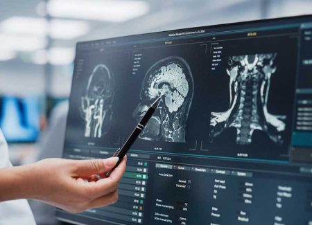
Diagnosis of cavernomas typically involves MRI or CT scans. MRI provides detailed images of vascular structures and surrounding tissues, helping determine the location and size of the cavernoma. In rare cases, other detailed imaging methods such as angiography may be used for differential diagnosis.
Treatment Methods
Treatment of cavernomas varies depending on symptoms and the risk of bleeding:
Observation:
For asymptomatic or low-risk bleeding cavernomas, active monitoring and regular MRI scans are recommended.
Surgical Treatment:
Microsurgical Excision: Used particularly for cavernomas with a history of bleeding or causing seizures. This method aims to control symptoms and prevent recurrent bleeding.
Radiosurgery:
In cases where surgical intervention poses risks, techniques like Gamma Knife radiosurgery may be preferred. This method targets the cavernoma with high-dose radiation, potentially leading to shrinkage over time.
Medical Treatment:
Antiepileptic Drugs: Used for seizure control, and analgesics for headache management.
Risks and Warnings
Bleeding Risk: Due to the fragile walls of vessels within cavernomas, there is a risk of bleeding. These bleeds can range from mild to severe and life-threatening conditions.
Seizure Risk: Cavernomas located in the brain may trigger seizures, necessitating long-term medication use.
Surgical Risks: Standard surgical risks such as infection, bleeding, or nerve damage apply. Some cavernomas may be difficult to access, making complete removal challenging and increasing the risk of recurrent bleeding.
Cavernomas are generally benign vascular lesions that can occasionally lead to significant health issues. Effective management and appropriate treatment options can mitigate the risks posed by these lesions and improve the quality of life for patients. Treatment choices should be tailored to the location, size, and functional impact of the cavernoma. Patients should be educated about potential risks and benefits of treatment options and maintain regular health check-ups.

